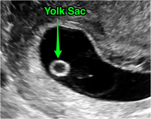
Gestational Sac And Yolk Sac. Two yolk sacs were identified in all but one case. The yolk sac is connected to the midsection of the embryos digestive tract by a narrow tube called the yolk stalk. The use of three-dimensional 3D ultrasonography has facilitated accurate volume estimation that has been confirmed in many organ. The ultrasound commonly shows a small collection of fluid within the lining of the uterus that represents the early development of the gestational sac.

It is visible at 5 weeks 5 days gestation. A fetal pole can be seen after the 6th week. The gestational sac may be recognized as early as 4 weeks and 1 day from the last menstrual period and should always be seen after 4 weeks and 4 days. Two yolk sacs were identified in all but one case. This is usually about five and a half weeks after a pregnant womans last period. The use of three-dimensional 3D ultrasonography has facilitated accurate volume estimation that has been confirmed in many organ.
Its IVF so I dont think dates can be wrong.
My repeat ultrasound is 8 days from now. The ultrasound typically shows a gestational sac and within it we can see a 3-5 mm bubble-like structure which is the yolk sac. The yolk sac nourishes the embryo and also helps produce blood cells during the early stages of pregnancy. Hi happymmummy I recently went for an emergency scan at 56 and saw a gestational sac and yolk sac but no baby. It is lined by extra-embryonic endoderm outside of which is a layer of extra-embryonic mesenchyme derived from. The yolk sac is surrounded by a larger black area known as the gestational sac.